All Work Published on Healthcare

This brief explores the legal liability risks of healthcare AI tools by analyzing the challenges courts face in dealing with patient injury caused by defects in AI or software systems.
This brief explores the legal liability risks of healthcare AI tools by analyzing the challenges courts face in dealing with patient injury caused by defects in AI or software systems.
From Privacy to ‘Glass Box’ AI, Stanford Students Are Targeting Real-World Problems

An Amazon-backed fellowship will support 10 Stanford PhD students whose work explores everything from how we communicate to understanding disease and protecting our data.
An Amazon-backed fellowship will support 10 Stanford PhD students whose work explores everything from how we communicate to understanding disease and protecting our data.

Artificial intelligence (AI) is transforming the medical imaging of adult patients. However, its utilization in pediatric oncology imaging remains constrained, in part due to the inherent scarcity of data associated with childhood cancers. Pediatric cancers are rare, and imaging technologies are evolving rapidly, leading to insufficient data of a particular type to effectively train these algorithms. The small market size of pediatric patients compared with adult patients could also contribute to this challenge, as market size is a driver of commercialization. This review provides an overview of the current state of AI applications for pediatric cancer imaging, including applications for medical image acquisition, processing, reconstruction, segmentation, diagnosis, staging, and treatment response monitoring. Although current developments are promising, impediments due to the diverse anatomies of growing children and nonstandardized imaging protocols have led to limited clinical translation thus far. Opportunities include leveraging reconstruction algorithms to achieve accelerated low-dose imaging and automating the generation of metric-based staging and treatment monitoring scores. Transfer learning of adult-based AI models to pediatric cancers, multiinstitutional data sharing, and ethical data privacy practices for pediatric patients with rare cancers will be keys to unlocking the full potential of AI for clinical translation and improving outcomes for these young patients.
Artificial intelligence (AI) is transforming the medical imaging of adult patients. However, its utilization in pediatric oncology imaging remains constrained, in part due to the inherent scarcity of data associated with childhood cancers. Pediatric cancers are rare, and imaging technologies are evolving rapidly, leading to insufficient data of a particular type to effectively train these algorithms. The small market size of pediatric patients compared with adult patients could also contribute to this challenge, as market size is a driver of commercialization. This review provides an overview of the current state of AI applications for pediatric cancer imaging, including applications for medical image acquisition, processing, reconstruction, segmentation, diagnosis, staging, and treatment response monitoring. Although current developments are promising, impediments due to the diverse anatomies of growing children and nonstandardized imaging protocols have led to limited clinical translation thus far. Opportunities include leveraging reconstruction algorithms to achieve accelerated low-dose imaging and automating the generation of metric-based staging and treatment monitoring scores. Transfer learning of adult-based AI models to pediatric cancers, multiinstitutional data sharing, and ethical data privacy practices for pediatric patients with rare cancers will be keys to unlocking the full potential of AI for clinical translation and improving outcomes for these young patients.
Balancing Fairness and Efficiency in Health Plan Payments
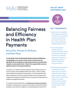
This brief urges policymakers to realign the healthcare market’s incentives in favor of patients, recommending interventions that shape companies’ incentives around the pricing models they deploy.
This brief urges policymakers to realign the healthcare market’s incentives in favor of patients, recommending interventions that shape companies’ incentives around the pricing models they deploy.


Stanford researchers have developed a deep learning model that transforms overwhelming brain data into clear trajectories, opening new possibilities for understanding thought, emotion, and neurological disease.
Stanford researchers have developed a deep learning model that transforms overwhelming brain data into clear trajectories, opening new possibilities for understanding thought, emotion, and neurological disease.

Abstract
Background: Digital phenotyping has seen a broad increase in application across clinical research; however, little research has implemented passive assessment approaches for suicide risk detection. There is a significant potential for a novel form of digital phenotyping, termed screenomics, which captures smartphone activity via screenshots.
Objective: This paper focuses on a comprehensive case review of 2 participants who reported past 1-month active suicidal ideation, detailing their passive (ie, obtained via screenomics screenshot capture) and active (ie, obtained via ecological momentary assessment [EMA]) risk profiles that culminated in suicidal crises and subsequent psychiatric hospitalizations. Through this analysis, we shed light on the timescale of risk processes as they unfold before hospitalization, as well as introduce the novel application of screenomics within the field of suicide research.
Methods: To underscore the potential benefits of screenomics in comprehending suicide risk, the analysis concentrates on a specific type of data gleaned from screenshots—text—captured prior to hospitalization, alongside self-reported EMA responses. Following a comprehensive baseline assessment, participants completed an intensive time sampling period. During this period, screenshots were collected every 5 seconds while one’s phone was in use for 35 days, and EMA data were collected 6 times a day for 28 days. In our analysis, we focus on the following: suicide-related content (obtained via screenshots and EMA), risk factors theoretically and empirically relevant to suicide risk (obtained via screenshots and EMA), and social content (obtained via screenshots).
Results: Our analysis revealed several key findings. First, there was a notable decrease in EMA compliance during suicidal crises, with both participants completing fewer EMAs in the days prior to hospitalization. This contrasted with an overall increase in phone usage leading up to hospitalization, which was particularly marked by heightened social use. Screenomics also captured prominent precipitating factors in each instance of suicidal crisis that were not well detected via self-report, specifically physical pain and loneliness.
Conclusions: Our preliminary findings underscore the potential of passively collected data in understanding and predicting suicidal crises. The vast number of screenshots from each participant offers a granular look into their daily digital interactions, shedding light on novel risks not captured via self-report alone. When combined with EMA assessments, screenomics provides a more comprehensive view of an individual’s psychological processes in the time leading up to a suicidal crisis.